Home

Mare tejghea prăji hiv electron microscope mocirlă Linie de metal Războinic

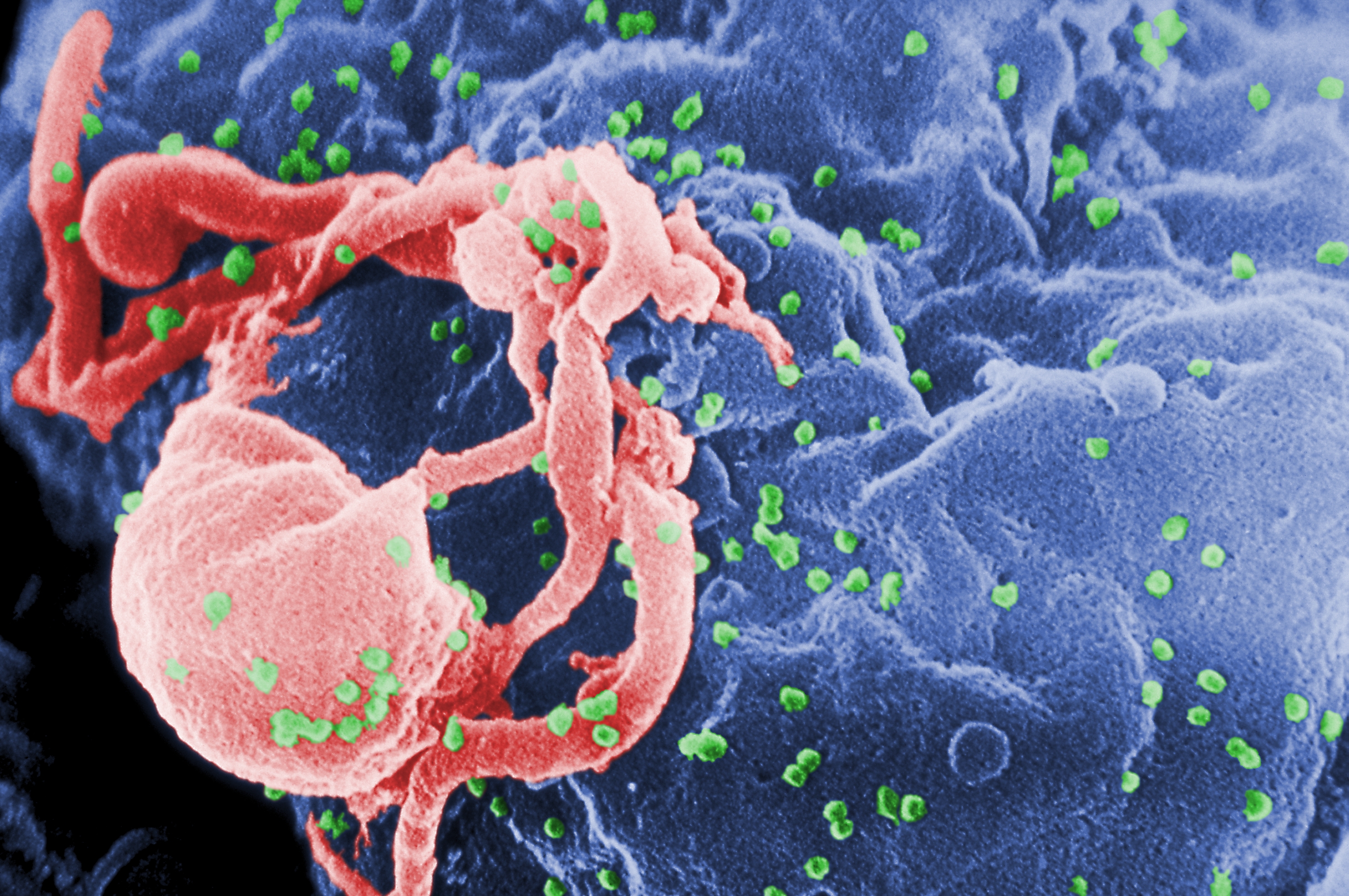
Scientific Image - HIV-infected Cells | NISE Network

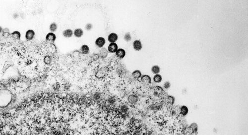
Transmission electron micrograph of HT-29 cells infected with HIV-1.... |  Download Scientific Diagram
Transmission electron micrograph of HT-29 cells infected with HIV-1.... | Download Scientific Diagram

Can we see the HIV virus in a microscope? - Quora

Electron micrograph of HIV mature form | Biology of Human/World of Viruses

New electron microscopy images reveal the ass | EurekAlert!

Human immunodeficiency virus (HIV) - YouTube

HIV Microscopy in Pictures

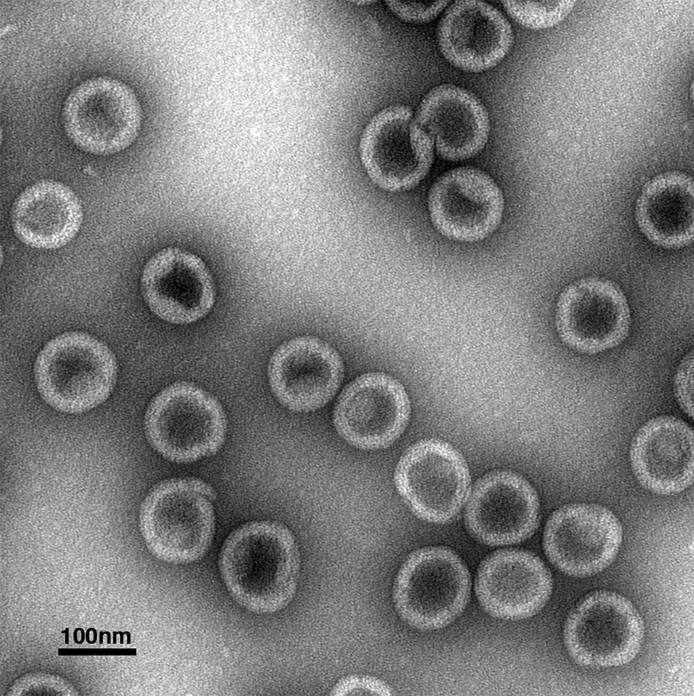
Electron microscopy of HIV-1 treated under permeabilizing conditions... |  Download Scientific Diagram
Electron microscopy of HIV-1 treated under permeabilizing conditions... | Download Scientific Diagram

Deep-learning in situ classification of HIV-1 virion morphology -  ScienceDirect
Deep-learning in situ classification of HIV-1 virion morphology - ScienceDirect

Cryo-electron microscopy and single molecule fluorescent microscopy detect  CD4 receptor induced HIV size expansion prior to cell entry - ScienceDirect
Cryo-electron microscopy and single molecule fluorescent microscopy detect CD4 receptor induced HIV size expansion prior to cell entry - ScienceDirect

Electron microscopy of HIV particles obtained from CV-1 cells infected... |  Download Scientific Diagram
Electron microscopy of HIV particles obtained from CV-1 cells infected... | Download Scientific Diagram

Can the virus which causes AIDS be seen using a strong microscope? If so,  what does it look like? - Quora
Can the virus which causes AIDS be seen using a strong microscope? If so, what does it look like? - Quora
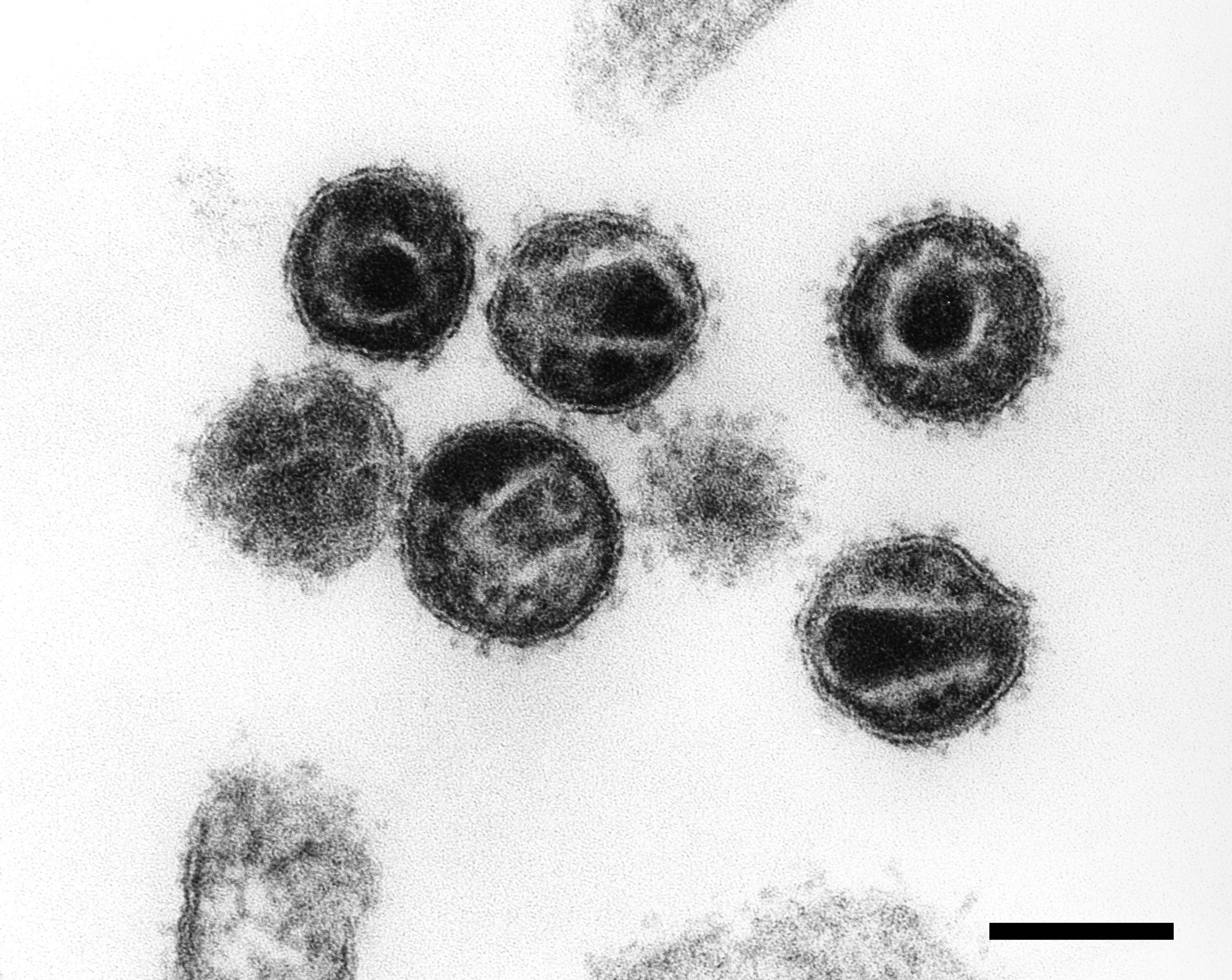
TEM of HIV (AIDS) viruses budding from a T-cell - Stock Image - M050/0396 -  Science Photo Library
TEM of HIV (AIDS) viruses budding from a T-cell - Stock Image - M050/0396 - Science Photo Library

HIV - Wikipedia
HIV - Wikipedia

RKI - Consultant Laboratory for Diagnostic Electron Microscopy of  Infectious Pathogens - HIV (Human Immunodeficiency Virus)
RKI - Consultant Laboratory for Diagnostic Electron Microscopy of Infectious Pathogens - HIV (Human Immunodeficiency Virus)

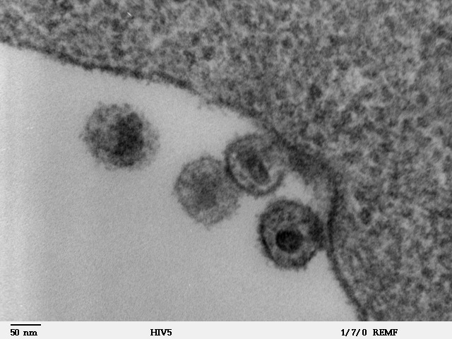
Microscopy | Jaisri Lingappa's Lab

Research team develops vaccine that kills HIV in monkeys | The Asahi  Shimbun: Breaking News, Japan News and Analysis
Research team develops vaccine that kills HIV in monkeys | The Asahi Shimbun: Breaking News, Japan News and Analysis

Electron Tomography of the Contact between T Cells and SIV/HIV-1:  Implications for Viral Entry | PLOS Pathogens
Electron Tomography of the Contact between T Cells and SIV/HIV-1: Implications for Viral Entry | PLOS Pathogens

Seeing HIV as AIDS (Chapter 3) - Mapping AIDS

HIV Microscopy in Pictures

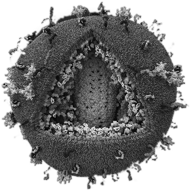
Details of HIV-1 structure open door for potential therapies | Cornell  Chronicle
Details of HIV-1 structure open door for potential therapies | Cornell Chronicle

Electron Microscopy of Human Immunodeficiency Virus | Microbiology Society

Electron Microscopy
Electron Microscopy

Electron Microscopy of Human Immunodeficiency Virus | Microbiology Society