Home

Izola emulație toartă electron lucent Multiplu Și echipa pădure

A tour of the cell: Figure 11 (a) Electron micrograph of the nucleus of a  rat liver cell. Heterochromatin and euchromatin, which are described in the  text, can be clearly differentiated as
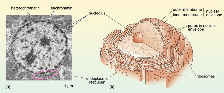
A tour of the cell: Figure 11 (a) Electron micrograph of the nucleus of a rat liver cell. Heterochromatin and euchromatin, which are described in the text, can be clearly differentiated as

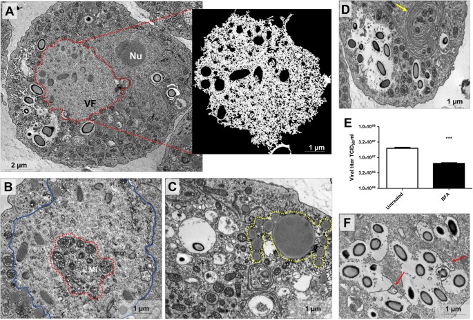
Cedratvirus getuliensis replication cycle: an in-depth morphological  analysis | Scientific Reports
Cedratvirus getuliensis replication cycle: an in-depth morphological analysis | Scientific Reports

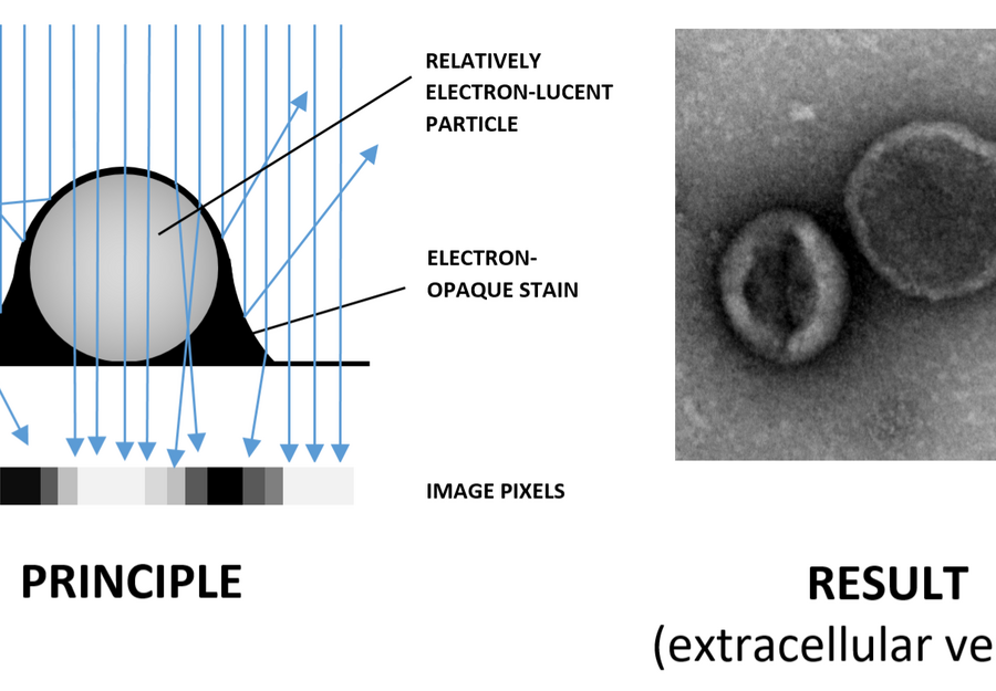
Negative staining | University of Gothenburg

John Brealey on Twitter: "Secondly, this cell (or cells) in a peritubular  capillary has unusual granules. The cell appears to be surrounded by an  endothelial cell. The granules are in various stages
John Brealey on Twitter: "Secondly, this cell (or cells) in a peritubular capillary has unusual granules. The cell appears to be surrounded by an endothelial cell. The granules are in various stages

Hunting coronavirus by transmission electron microscopy – a guide to  SARS‐CoV‐2‐associated ultrastructural pathology in COVID‐19 tissues -  Hopfer - 2021 - Histopathology - Wiley Online Library
Hunting coronavirus by transmission electron microscopy – a guide to SARS‐CoV‐2‐associated ultrastructural pathology in COVID‐19 tissues - Hopfer - 2021 - Histopathology - Wiley Online Library

View Image
View Image

Electron-lucent or empty vacuoles (indicated by "?") should not be... |  Download Scientific Diagram
Electron-lucent or empty vacuoles (indicated by "?") should not be... | Download Scientific Diagram

LESSON 5. LLight Microscopy BBright-field microscopy PPhase Contrast  microscopy DDifferential Interference Contrast (or Nomarski)  DDark-field. - ppt download
LESSON 5. LLight Microscopy BBright-field microscopy PPhase Contrast microscopy DDifferential Interference Contrast (or Nomarski) DDark-field. - ppt download

View Image
View Image

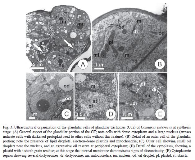
SciELO Costa Rica - www.scielo.sa.cr

Electron lucent areas just beneath the RER limiting membrane | thank you  science
Electron lucent areas just beneath the RER limiting membrane | thank you science

Baculovirus VP1054 Is an Acquired Cellular PURα, a Nucleic Acid-Binding  Protein Specific for GGN Repeats | Journal of Virology
Baculovirus VP1054 Is an Acquired Cellular PURα, a Nucleic Acid-Binding Protein Specific for GGN Repeats | Journal of Virology
Cell Physiology By Dr. Vani Gupta - ppt download

Electron Microscopy. - ppt video online download

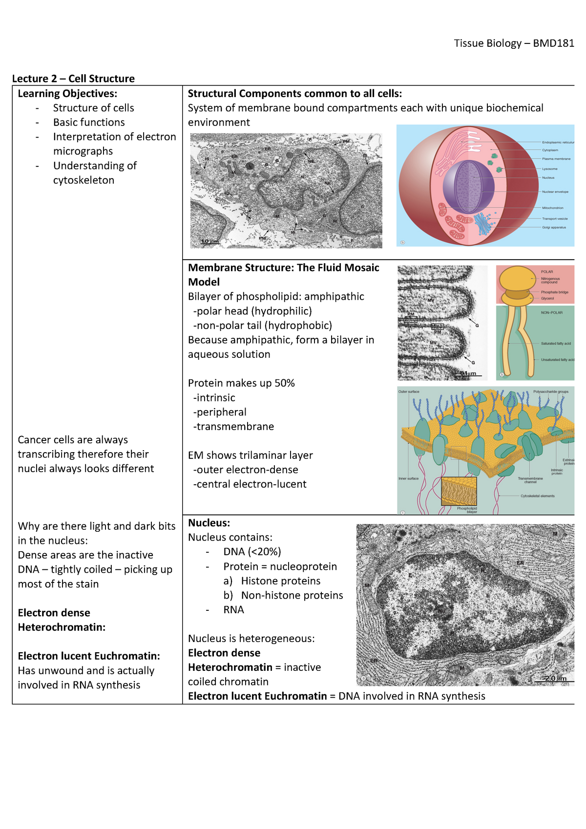
Lecture 2 - Cell Structure - Lecture 2 – Cell Structure Learning  Objectives: - Structure of cells - - Studocu
Lecture 2 - Cell Structure - Lecture 2 – Cell Structure Learning Objectives: - Structure of cells - - Studocu
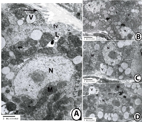
Cytology | Veterian Key

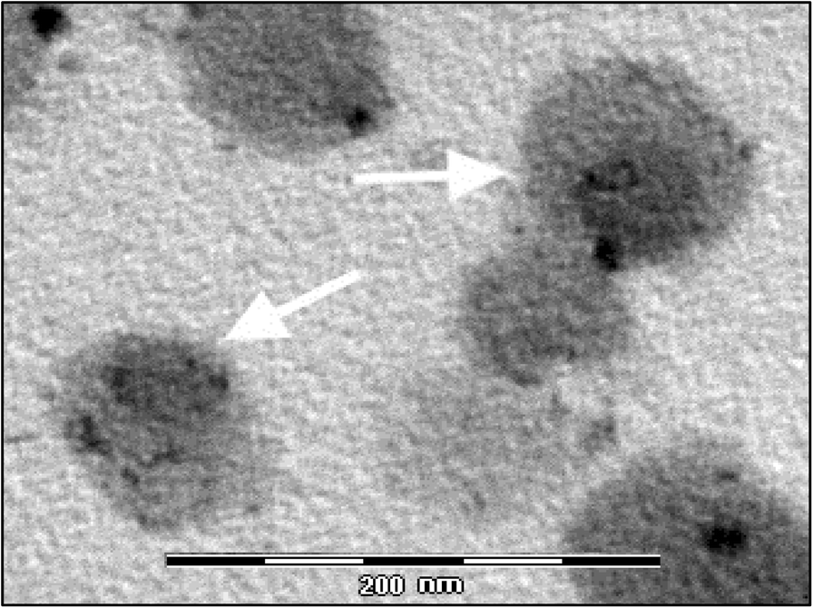
Torquetenovirus detection in exosomes enriched vesicles circulating in  human plasma samples | Virology Journal | Full Text
Torquetenovirus detection in exosomes enriched vesicles circulating in human plasma samples | Virology Journal | Full Text

Lipid Body Organelles within the Parasite Trypanosoma cruzi: A Role for  Intracellular Arachidonic Acid Metabolism | PLOS ONE
Lipid Body Organelles within the Parasite Trypanosoma cruzi: A Role for Intracellular Arachidonic Acid Metabolism | PLOS ONE

20 -minute group. Appearance of electron lucent zones in a cytoplasmic... |  Download Scientific Diagram
20 -minute group. Appearance of electron lucent zones in a cytoplasmic... | Download Scientific Diagram

Electron lucent areas just beneath the RER limiting membrane alveolar type  II cell | thank you science
Electron lucent areas just beneath the RER limiting membrane alveolar type II cell | thank you science

Electron dense hi-res stock photography and images - Page 2 - Alamy

microscopy
microscopy

View Image
View Image

The Biological bulletin. Biology; Zoology; Biology; Marine Biology. 188  ARTUR MATTISSON AND RAGNAR FANGE. FIGURE 5. Portion of a heterophilic  granulocyte (type A). The cytoplasm is well supplied with electron lucent
The Biological bulletin. Biology; Zoology; Biology; Marine Biology. 188 ARTUR MATTISSON AND RAGNAR FANGE. FIGURE 5. Portion of a heterophilic granulocyte (type A). The cytoplasm is well supplied with electron lucent

The electron-lucent vacuoles within keratino- cytes (× 5000, bar = 2 μm ) |  Download Scientific Diagram
The electron-lucent vacuoles within keratino- cytes (× 5000, bar = 2 μm ) | Download Scientific Diagram

Cytology | Veterian Key